In childhood, emergency dental care is essential to good health. The preservation of deciduous teeth with injured pulp by decay or trauma can be a major problem. Dentistry has been searching for decades for an effective method of treatment. There have been proposed many techniques. The dentist will recognize familiar names such as direct pulp capping, indirect pulp capping, partial pulpotomy, pulpotomy and pulpectomy. They have advised different drugs and medication to follow these techniques, and there have been reports of varying degrees of success. Unfortunately, many of these techniques have been subject to controversy and the results are unpredictable.
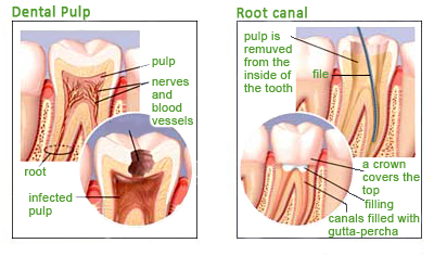
The dental pulp contains elements that make it similar to other loose connective tissues. Within the pulp are blood vessels, lymph vessels, nerves, defenses cells, ground substance and fibroblasts. Yet another feature is the presence of pulp odontoblasts, necessary for the production of dentin.
From the point of view of development, the dental pulp emerges as a result of the promotion of the dental lamina of mesoderm to form the dental papilla. Its shape is determined by the enamel organ. When this mature embryonic tissue, dentin deposited odontoblasts in cusp tips are formed. When mature the dental papilla creates dentin and directed distally, and the tissue becomes more cellular and vascular.
Each element in the structure of the dental pulp plays an important role in the life and preservation of the piece. Fibroblasts produce tropocollagen, which in turn is converted to collagen fibers. The base substance binds the fibers together. Its chemical action plays an important role during inflammation. Odontoblasts, which evolves dentin creates a cell cytoplasm is evident not only in the pulp, but also on dentine.
The odontoblasts are seen as cells with long extensions that intertwine and become even more profuse when approaching the junction between enamel and dentin. The pulp also contains undifferentiated mesenchyme cell that can develop into odontoblasts, histiocytes act as phagocytes, and wandering lymphatic cells operating in the production of antibodies. In each tooth pulp there is an intricate arrangement of arteries and veins that in turn communicate with the rest of the body. Similarly, there is a lymphatic network functions similarly to that in other areas of the body. Autonomic and sensory nerves complete the items “bind” to the body part.
For the transmission of stimuli to the autonomous capillary vasodilation creates increased pressure on the nerve endings of free or sensory nerves and in turn a reaction of pain is experienced.
The dental pulp and its physiological functions are similar in many respects to other body parts. However, their individual characteristics such as large confinement structurally hard dentin have a unique situation. A responsible clinician operator must know the structure of the pulp and be aware of the limitations of their treatment to achieve optimal results in the treatment of the diseased or traumatized parts.